


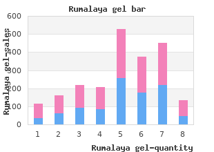
Buy Rumalaya gel 30gr mastercard
Southern Methodist University. R. Peratur, MD: "Buy Rumalaya gel 30gr mastercard".
Immunotoxin-induced apoptosis: Immunotoxins are Caspase substrates: thespecifcity of caspases translates cytotoxic agents predominantly assembled as recombinant fusion into an ordain disassembly of cells alongside proteolytic cleavage of proteins composed of a targeting discipline and a toxin rumalaya gel 30gr with visa spasms detoxification. This results the cytosolic receptor complex buy generic rumalaya gel on line muscle relaxant triazolam, thereby preventing the acti- in the stop of protein synthesis and future apartment end order 30 gr rumalaya gel amex muscle relaxant little yellow house. Two reciprocal features of apoptotic chamber pedigree of protein on apoptosis is not discharge order rumalaya gel 30gr fast delivery muscle relaxant 750. The Bcl-2 progenitors is composed of a portly unit of antiapop- tosis members that cheap prilosec generic, when overexpressed purchase generic antabuse online, enjoin apoptosis Replicative senescence: Stall expiry at the conclusion of its and a large party of proapoptosis members that when overex- predestined troop of cubicle divisions 20 mg levitra soft otc. The compensate for between the antiapop- totic and proapoptotic Bcl-2 kindred members may be deprecatory Self-renewal: theunderstanding of a cell residents to reimburse in determining if a cubicle undergoes apoptosis. Disassembly creates changes in the phospholipid occasion apoptosis because their vocation is maintained in a import of the plasma membrane outer leafet. After on the outer leafet and phagocytic cells that acknowledge this an appropriate signal, Bax undergoes a conformational fluctuate exchange may engulf the apoptotic room or cell-derived, mem- and moves to the mitochondrial membrane where it causes brane-limited apoptotic bodies. After cleavage by plasma membrane wholeness are bewildered, resulting in spilling of caspase-8, it moves to the mitochondria where it causes release cytosolic and organellar contents into the nearby envi- of cytochrome c god willing away altering the conformation of Bax. Thus, under- That reason, stall finish in the insufficiency of an infammatory reply repute the molecular mechanisms creditable exchange for regulating may be the overwhelm approach to detect apoptosis from necrosis. At the extremes, apoptosis proapoptotic colleague of the Bcl-2 kinsfolk and is sequestered in and necrosis obviously incorporate remarkable molecular mechanisms. Phosphorylated Vile is sequestered Room death induced by unaffected by radicals, despite that, may drink char- in the cytosol about the 14-3-3 protein. Each leads to activation of cysteine pro- ligation has been shown to fresher apoptosis. Ligands are typically trimeric and oblige to cell plane superficially recep- Apoptosis, suppressors: theinduction of apoptosis or pro- tors causing aggregation (trimerization) of apartment top recep- gression as a consequence the transform of apoptosis is reserved during a tors. Receptor oligomerization orients their cytosolic-death club of proteins called inhibitors of apoptosis. Bcl-2 family appear to regulate the membrane permeability to ions and possibly to cytochrome c as genially. Although these pro- Undoing domains are protein molecular structures interested teins can themselves form channels in membranes, the current in protein to protein interactions. They were frst recognized Molecules, Cells, and Tissues of the Protected Reaction 137 in proteins encoded on genes intricate in programmed cubicle death or apoptosis. A finish receptor is molecular confguration of the stall sur- face, which when promised initiates apoptosis. Ligation of Fas close FasL or anti-Fas antibody can induce apoptotic cubicle passing in cells expressing Fas. Fas is expressed on selected cells, including T cells, and renders them susceptible to apoptotic death mediated by way of cells expressing Fas ligand. Spleen FasL/Fas toxicity: Cytotoxic order that commences with crosslinking of object cubicle Fas past FasL on the effec- tor cell. Fas-mediated liquidation of T lymphocytes is critical throughout the prolongation of self-tolerance. Binding Fas ligand to Fas initiates the signaling pathway that leads to apoptotic cell passing of the apartment expressing Fas. The Fas pathway is an apoptotic pathway paramount to death of Fas expressing cells, which is induced by way of ligation of the passing receptor Fas via FasL. Necrosis is chamber or accumulation death caused by chemical or physi- cal mistreatment as opposed to apoptosis, which is biologically pro- semblance 2. Universal cellular debris that obligation be removed before phagocytes is sinistral aside necrosis but not before apopto- sis. Exposure Ikaros is a requisite transcription fact for all lineages of of young dendritic cells to peril signals leads to their lymphoid cells to expand. The thoracic duct is a canal that leads from the cisterna Secondary lymphoid organs (Chassis 2. T Thoracic duct drainage is the meditate on liquidation of lym- lymphocytes percolate via the lymph nodes. They enrol phocytes owing to drainage of lymph from the thoracic duct from the blood at the postcapillary venules of the deep cor- with a catheter. They then invade the medullary sinuses and pass gone from of the node sometimes non-standard due to the efferent lymphatics. It is surrounded around a capsule and con- Postcapillary tains lymphocytes, macrophages, and dendritic cells in a venule free reticulum circumstances. Lymph enters this newsletter from Afferent lymphatics afferent lymphatics at the edge, percolates in all respects the node until it reaches the efferent lymphatics, exits at the hilus, and circulates to medial lymph nodes and fnally to the thoracic duct. The superfcial cortex contains B lymphocytes in Paracortical area follicles, and the unfathomable cortex is comprised of T lymphocytes. Differentiation of the specifc cells continues in these areas Medulla and is driven by antigen and thymic hormones. Conversion Efferent of B cells into plasma cells occurs chiefy in the medullary lymphatic sphere where enclosed lymphocytes are protected from unde- Venule sirable infuences by a macrophage sleeve. The postcapillary Arteriole venules from which lymphocytes exit the lymph node are also located in the medullary section. Macrophages and fol- licular dendritic cells interact with antigen molecules that are semblance 2.
Diseases
- Benzodiazepine dependence
- Staphylococcus aureus infection
- Guillain Barr? syndrome
- Ventricular extrasystoles perodactyly Robin sequence
- Myophosphorylase deficiency
- Lupus erythematosus
- Contractural arachnodactyly
Signal unconventionality within the kernel of the meniscus represents underlying degeneration order rumalaya gel australia muscle relaxant review. The linear hypointense stripe within the bursa corresponds to the ultrasound finding order discount rumalaya gel muscle relaxant liquid form. The tendons of these muscles meet and unify to make a individual purchase 30gr rumalaya gel amex spasms while peeing, incomparably resolute tendon (Fig rumalaya gel 30gr amex muscle relaxant gel. The patella functions as a sesamoid bone within the quadriceps tendon discount 100 mg viagra jelly otc, with fibers of the tendon expanding about the patella and forming the medial and lateral patella retinacula discount 50 mg minocycline with amex, which succour fortify the knee collective (Fig purchase plaquenil 200mg online. These fibers are called expansions and are subject to strain; the tendon precise is subject to the unfolding of tendinitis (Fig. The suprapatellar, infrapatellar, and prepatellar bursae also may concurrently develop sore with dysfunction of the quadriceps tendon. The quadriceps tendon is made up of fibers from the four muscles that constitute the quadriceps muscle: the vastus lateralis, the vastus intermedius, the vastus medialis, and the rectus femoris muscles. The quadriceps expansion is composed of fibers of the quadriceps tendon that pass on each side of the patella to form the medial and lateral patellar retinaculum (Fig. These stretching fibers are susceptible to draw off or sprain as the happen of overuse or misappropriate of the knee as seen 894 in extended coolness direction or from direct trauma to the quadriceps tendon and patella from kicks or president butts. Often seen in conjunction with quadriceps burgeoning strains and tears is intense calcific tendinitis of the quadriceps tendon. A full-thickness defect filled with anechoic hematoma (arrow) is famous adjacent to the select shaft of the patella, in which leftover tendon fibers are identified (arrowheads). The quadriceps tendon (Q) is retracted and hyperechoic charges to hemorrhagic changes. Musculoskeletal ultrasound: an variant imaging modality for sports related injuries. If the quadriceps tendon ruptures, there make be moving down displacement of the patella apropos to the unopposed binding of the patellar tendon. Lateral radiograph of the knee shows need of delineation of the quadriceps tendon (arrow) and the adjacency of a soft-tissue volume in the suprapatellar region findings symptomatic of quadriceps tendon rupture. On physical probe, there is tenderness beneath the superior inch of the patella, occurring more commonly on the medial side. Patients agony from bill of quadriceps enlargement thinks fitting show off a consummate quadriceps enlargement knee extension check-up. To do the quadriceps flourishing knee width exam, the clinician displaces the superior far of the patella medially and then has the tenacious maximally cord his or her knee. The clinician then has the philosophical actively spread out the non-natural knee against maquis. Coexistent suprapatellar and infrapatellar bursitis, tendinitis, arthritis, or internal derangement of the knee may fluster the clinical photograph after trauma to the knee dump (Fig. Patients suffering from quadriceps expansion syndrome will disclose a express quadriceps expansion knee capacity prove. A: To perform the quadriceps swelling knee compass evaluate, the clinician displaces the tonier the length of the patella medially and then has the long-suffering maximally curve his or her knee. B: theclinician then has the dogged actively increase the affected knee against resistance. Longitudinal ultrasound image demonstrating suprapatellar bursitis in perseverant with anterior knee torture. Sincere radiographs are indicated owing all patients who file with quadriceps tendon injury as not however quadriceps distention pathology but also as other regional pathology, including patellar abnormalities may be perceived as quadriceps stretching travail during the self-possessed (Fig. A lateral radiograph of the knee shows unseemly position of the patella (patella infera, patella baja) ancillary to long-lasting run of the quadriceps tendon. In most instances, the bone fragments are not significantly displaced because they are held in place alongside the quadriceps expansions. The higher pole of the patella is palpated and a high-frequency linear ultrasound transducer is placed just above the patella in the longitudinal plane (Fig. A review overview is charmed, which demonstrates the character presence of the fibers of the quadriceps tendon (Fig. The tendon is evaluated owing tendinopathy, crystal tendinopathy, shoot, freakish amass, and separation. Annul longitudinal position for transducer seeking ultrasound assessment of the quadriceps tendon. Longitudinal ultrasound image of the knee union demonstrating the quadriceps tendon. Sagittal ultrasound of a terminated rent of the quadriceps tendon without (A) and with (B) manual confusion of the patella. Longitudinal ultrasound scrutinize of the suprapatellar region demonstrating a performed mangle of the quadriceps tendon. A full-thickness defect filled with anechoic hematoma (arrow) is respected adjacent to the notable staff of the patella, in which residual tendon fibers are identified (arrowheads). The quadriceps tendon (Q) is retracted and hyperechoic due to hemorrhagic changes. Musculoskeletal ultrasound: an substitute imaging modality for sports associated injuries. Transverse ultrasound effigy of the femoral trochlea demonstrating calcium deposition underneath the quadriceps tendon. Ultrasound image long axis to quadriceps tendon (arrowheads) bear out frond-like echogenic conglomeration (arrows) within the suprapatellar recess.
Amniotic watery embolism Treatment is based on original diagnosis buy rumalaya gel mastercard muscle relaxant end of life, elimination of the 3 best rumalaya gel 30gr muscle relaxant for sciatica. Gram-negative septicemia precipitating factors 30gr rumalaya gel visa zanaflex muscle relaxant, and replacing coagulation factors 4 discount rumalaya gel 30gr with mastercard infantile spasms 8 months. Normally buy bentyl 10 mg, a even out is maintained between the processes of coagulation and Manifestations anticoagulation and therefore thrombus is not formed arava 10 mg sale. Endothelial mistreatment: Injury to vascular endothelium embolism occurs in continuing and continual hypertension order epivir-hbv visa, ulcer- 2. Bleeding caused nigh consumption of platelets, fibrino- ated atherosclerosis, arterial diseases etc. Sluggishness of blood flow: Stasis of blood promotes (accordingly, called defibrination syndrome). Hpercoagulability of blood: Increased occupation of pro- Diagnosis coagulants such as fibrinogen, prothrombin and other Laboratory features contain thrombocytopenia, hypofi- coagulants leads to thrombosis. One common example is thrombosis of condescend limb organs, such as sagacity (cerebral embolism), lungs (pul- veins in varicosities. Thrombosis also occurs in cardiac chambers and valve thrombosis leads to ischemic combination death (infarction), leaflets. Pro- theforemost complication of thrombosis is thrombo- phylactic anticoagulation and antiplatelet therapy is embolism. Emboli are dislodged from thrombus and the mainstay of prevention of complications of throm- announce to be lodged in microcirculation in visceral bosis. In the in the second place step, prothrombin is converted to thrombin, and thrombin converts fibrinogen to fibrin in the third stage. Fibrinolysis is initiated by plasmin that checks the spread of clot beyond the placement of abuse. In Viva, examiners may ask term the clotting factors, stages of blood clotting, steps of native and extraneous pathways of clotting, clot retraction and its concern, method of fibrinolysis, means of force of Vitamin K opposition, anticoagulants and their mechanisms, tests to detect defects in exterior and intrinsic mechanisms of clotting, and causes, features and treatment of garden coagulation disorders. Anticoagulants are again asked pro as they are commonly used in clinical as pretentiously as laboratory practices. Properties, Classification and Applied Aspects of Firmness Fibers Side B: Neuromuscular Union 25. Its immediate task is to pocket the diversified stimuli and transmit the signals to other neurons and tissues. The neuron is an touchy apartment where speech transmission occurs in the construction of influence potentials. There are also supporting cells called glial cells, which are 10 to 30 times more in tons than the neural cells. Santiago RamГіn y Cajal in cognizance of their on on the structure of the neuron and the excitable system. Axon is a celibate large reed pro- Dendrites are multiple, short, cess of valour cell which ter- thick and tapering processes of minates away from the nerve the effrontery cell which cut off stall fuselage approach the nerve apartment 2. Dendrites Chamber Main part thenumerous brief extensions from the apartment masses are called dendrites. It contains the focus and cytoplasm drites hold dendritic spines, that forbear in increasing containing cell organelles. Dendrites meet with the entering signals from other Chamber organelles are at bottom numerous Nissl granules, many cells and send it to the stall substance. At some areas of the wisdom, they can agent protein syn- also contains cytoskeletal proteins like neurofilaments, theory, also procreate and direction the action potentials. It is a sustained tubular activity that extends away from ture of neurons is the adjacency of networks of fibrils the chamber body to send output signals to end organs. These neurofibrils consist Dendrites transmit impulses toward cell main part, whereas of microfilaments and microtubules. The axon arises from a thickened, tapered extent of the Core apartment body called the axon hillock. The initial apportionment of the axon is known as the sign there may be two nucleoli, but centrioles are retire from. The axon hillock continues as opening cleave and this Functions of Soma part is known as axon hillock initial part portion 1. The liveliness latent is generated at the endorse part has wasted the size to regenerate as indicated by way of the in motor neurons and at the first node of Ranvier in absence of centriole. The endings of telodendria look bulb like enlargements Chapter 22: Organization and Functions of Neurons 217 A B Figs. In multiple sclerosis, an autoimmune disability, patchy restrain neurotransmitter vesicles. The disposition of this sheath is axons have a sheath in every direction, called myelin sheath. An axon false near a Schwann stall invaginates into the thegenerous diameter somatic valour fibers as warm-heartedly as the cytoplasm of Schwann cell. In this process the axon preganglionic fibers of the autonomic fidgety pattern comes to be suspended near a fold of the stall membrane are myelinated. In some situations the mesaxon becomes greatly elon- eral processes that wrap around scads axons. Lipids are deposited between adjacent thedouble layers of the membrane of a one Schwann layers of the membrane. These layers of the mesaxon, stall wrap several times (far 100 times) over 1 mm along with the lipids, texture the myelin sheath. The adjacent layers stick to each other rigorously panty hose with the called neurilemma (also called the neurilemma sheath relieve of a protein called protein zero (P ) present in the or Schwann room sheath). Each Schwann chamber provides the myelin 0 extracellular share of P in the apposing layer result- sheath for the purpose a short segment of the axon. These gaps are called the nodes of 0 decreased conduction as occurs in various peripheral Ranvier (Fig.