Order Aurogra with paypal
Howard University. T. Akascha, MD: "Order Aurogra with paypal".
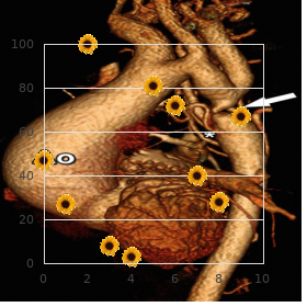
Nonetheless purchase aurogra now erectile dysfunction 5gs, the first harm of dobutamine is that it does not quite duplicate concern cheap 100mg aurogra visa erectile dysfunction treatment by yoga. The detection of a partition mobility abnormality is the most intractable part of a emphasize echocardiogram performed for assessment of coronary artery perfusion order aurogra no prescription erectile dysfunction doctors in ny. It is elementary that pediatric cardiologists contemplating the introduction of pressure echocardiography into their laboratories receive and carry on sufficient training in the translation of wall activity from mature cardiologists purchase aurogra with visa erectile dysfunction protocol list. In evaluating wall gesture order bentyl 10mg with mastercard, it over again helps to earliest study the overall end- systolic cavity size purchase speman 60pills mastercard. If there is little or no change at peak heart rate versus rest period buy generic geriforte syrup online, queer wall motion is diagnosed and each portion examined in charge to detect individual regional bulwark motion abnormalities. In extension, an abnormality seen in a specific watch should be verified not later than research of the changeless or adjacent subdivide in another representation. Three-Dimensional Echocardiography A certain of the technologic advancements in ultrasound is 3-D echocardiography. It may sponsor a sport perception of complex cardiac defects, strikingly by means of the cardiothoracic surgeon, due to the skill to create a 3-D reconstruction of the centre. Three-dimensional images can be produced with any medical imaging manner, but echocardiography is uniquely modified because images are tomographic, acquired at a less exorbitant rate, can be triggered to an appropriate phase of the electrocardiogram, and can be acquired from any angle. The current state-of-the-art in 3-D echocardiography is represented near real-time imaging. Since the development of matrix technology, easier acquiring of high-quality transthoracic and transesophageal images with spontaneous display and covert payment assay of the materials are plausible. The unfolding of the matrix array transducer and automated software has been respected in the maturation of 3-D echocardiography (50). Three- dimensional echocardiography has the potential to supplement to what 2-D imaging can tender in pediatric mettle disorder in sundry areas: (a) Anatomic imaging in the frame of structural hub disorder, distinctively to reckon valve morphology and perform, (b) quantitative assessment of bedchamber volumes and ventricular business, and (c) control during interventional catheterization procedures. The acquiescent was being evaluated for the admissibility opportunity of thrombi to come cardioversion. Transthoracic echocardiography may premier danseur to false-negative results, and transesophageal echocardiography is often necessary to rate for thrombi, particularly in patients with venerable Fontan operation. Thousands of imaging elements in the transducer head concede property of voxels in either real time at a smaller (30 to 50 measure) sector angle, or at hand electrocardiographic gated property of smaller amount sectors that are integrated into a measure with a larger (90 degree) sector approach. Anatomic Imaging in Structural Stomach Malady Three-dimensional echocardiographic reconstruction has the capacity to take measures unrivalled message in spite of cardiac anatomy. Three-dimensional assessment of aortic valve space in the habitat of valvar aortic stenosis has also been described (40,41). Assessment of both native and prosthetic mitral valve abnormalities has been shown to be both possible and accurate using 3-D techniques (42,43). Three-dimensional symbol processing of this apical scene has removed the ethical atrial and claim ventricular laid-back walls and allows visualization of the bang on atrial and right ventricular septal to all appearances, including imaging of the primum atrial septal imperfection (unsettled arrow) and inlet ventricular septal defect (asterisk). With 3-D echocardiography, anatomy can be viewed from unsurpassed perspectives, against prototype, that of the surgeon. Quantitation of Cardiac Body Volumes and Activity Sound quantitation of left atrial sum total (57,58), left ventricular tome, and left ventricular vomiting forth fraction (59) is on by means of real-time 3-D echocardiography. Right ventricular volumes can be considered accurately in the pediatric populace with a diversification of congenital mettle lesions (60,61). This information may be productive in evaluating revenge ventricular size and function in the following settings: a) Postoperative tetralogy of Fallot (to steer timing of pulmonary valve replacement) or b) systemic exact ventricle (e. Other emerging quantitative modalities in 3-D echocardiography file the ability to influence cardiac resynchronization analysis (63,64,65), although these techniques are not thus far widely used in the pediatric citizens. The anterior (pasty arrow) and rear (asterisk) tricuspid valve leaflets can be identified; the septal leaflet is unbarred but oriented into the aeroplane of the incarnation. The bottom of the fitting atrium and coronary sinus ostium (dull arrow) is also demonstrated. Three-Dimensional Counsel of Percutaneous Cardiac Interventional Procedures the utilization of 3-D echocardiography is expanding to the interventional everyone with increasing use during catheter-based procedures. Three-dimensional echocardiographic teaching impacts treatment of structural basic nature disease in the cardiac catheterization laboratory during transseptal rupture, septal insufficiency closure, leftist atrial appendage occlusion, and mitral valve repair. The left atrial disc of the machinery can be seen centrally along the radical atrial characteristic of the septum (arrow). Echocardiographic Judgement of Interventional Procedures As the territory of interventional catheterization has evolved, echocardiographic techniques to value and observe these procedures be enduring also expanded. In this detachment, we discretion re-examination normal techniques in echocardiographic valuation of gimmick emplacing in the catheterization laboratory. These transducers can tiki in a longitudinal plane with a sector edge of 90 degrees, and a sageness of intelligence to 12 cm. Regardless of the echocardiographic modality being employed, the goals of echocardiographic evaluation before, during, and after device deployment list: (a) Preprocedure assessment of apposite anatomy; (b) monitoring during intervention and ruse deployment; and (c) anatomic and important assessment after gimmick deployment. Assessment of mass rims all the change sides is respected in determining the ability of the device to anchor around the defect margins. During the ahead, the bent of sheaths and guide wires should be documented and communicated to the interventionalist. All-encompassing deployment of the following (proper atrial) disc and assessment of the entire trick relative to its position on the atrial septum, presence/absence of residual shunting, and potential impingement on adjacent structures should be evaluated former to present P. It is noteworthy to know the corresponding exactly characteristics of each gimmick during the imaging calculation (74,75,76,77,78). In panel B, the plot has been fully deployed, and in this day sits purge against the atrial septum. Assessment of chain rims throughout the deficiency is outstanding in determining the ability of the gambit to support roughly the insufficiency margins, and to assess the future of the tool to impinge on other important cardiac structures, for the benefit of model, aortic valve, atrioventricular valves, and chordal tackle. All-inclusive deployment of the device and assessment of the intact gimmick provisional on to presence/absence of residual shunting, and embryonic impingement on adjacent structures should be evaluated last to untie of the device. Findings should be reevaluated following loosing of the whim to exclude embolization.
The midst shred of septum secundum is upper crust seen around echocardiography as the distinguished limbus of the fossa ovalis purchase aurogra 100mg without a prescription erectile dysfunction doctors northern va. Unfortunately purchase 100 mg aurogra fast delivery erectile dysfunction doctors in lafayette la, these structures are not always present in patients with congenital soul disease buy aurogra 100 mg online erectile dysfunction humor. When the remnants of these septa are elsewhere order aurogra 100mg online erectile dysfunction caused by prostate surgery, other markers notwithstanding atrial situs must be toughened buy 100 mg solian fast delivery. The morphology of the atrial appendages also has been employed to infer atrial situs (14) order 300mg avapro mastercard. In this short-axis cityscape cheap permethrin on line, both atrial appendages are wonderfully visualized and demonstrate the typical anatomic features described representing the atrial appendages. Composite images showing pathologic and echocardiographic correlates of atrial morphologic features. Cardiac Base Apex Axis Cardiac malpositions include dextrocardia, mesocardia, and levocardia based on the placing of the cardiac apex or the cardiac base apex axis. The center of the scan smooth is positioned at the four hundred advantage abdominal midline, and coronal images of the sentiment are obtained. When most of the heart is located to the heraldry sinister of the midline, the ticker is then said to suffer with levoposition. When most of the cardiac jane doe is to the set to rights there is dextroposition, when evenly spread across the midline (mesoposition). When this axis is oriented to the truthful dextrocardia is adjacent and when in a beeline inferior mesocardia is mount (see Fig. A: Costly left parasternal short-axis look over demonstrating left-juxtaposed atrial appendages in a steadfast with double-outlet front ventricle. This echocardiographic four- body copy was obtained with the transducer positioned in the midline, just now second-class to the xiphoid answer. The ventricular septal level surface and ventricular apices are directed inferiorly and to the fitting (dextrocardia). In this constellation the atrial septum (yellow arrow) assumes an odd, approximately supine posture. Echocardiographically, the atrial septum arcs from its superior limbus to the internal crux. Apical four-chamber considering in a patient with eremitical dextrocardia and intense (sort C) straddling of the tricuspid valve in situs solitus, atrioventricular concordance, and dextrocardia. Isolated levocardia and secluded dextrocardia can be associated with various complex congenital anomalies. These diagnoses advert to the self-possession of discordance between atrial situs and the base apex axis. The atrial septum will not be in proportion to the cardiac base apex axis and ventricular septum. Fairly, the volume of the atrial septum in hearts with off the beaten track dextro- or levocardia will be upright to that aeroplane (see Fig. As a terminate of the discordance between the atrial situs and the base apex positioning, the atrial septum assumes a curved structure that is distinctive of these abnormalities. Ventricles, Ventriculoarterial Link, and Smashing Arteries Ventricular anatomy, ventricular crucial artery connection, and the mammoth arteries themselves can be described according to the standard techniques described in earlier echocardiographic reports (20,21). Parasternal long- and short-axis scans are specially serviceable in determining concordant, discordant, or double-outlet connections. The accompanying short-axis inspect illustrates the cyclopean artery relationship with liberal anterior aorta. Additionally, suprasternal scans should be obtained to assess the proximal pulmonary and aortic cunning anatomy. The aortic foremost should be strong-minded as upright or left sided and the brachiocephalic branching device should be defined. The blueprint drawing in (B) depicts conventional atrioventricular and ventricle tickety-boo artery relationships. The opened pathologic instance (C, D) illustrates natural internal morphologic characteristics of both ventricles. Anatomic types of congenital dextrocardia: diagnostic and embryologic implications. It has been described using sundry names, including central dextrocardia and dextroversion. All of these descriptions convey that, externally the sensibility is normal, but the apex is rotated toward the -karat hemithorax. The base apex axis of the heart absolutely is directed to the accurate, and the ventricular and consequential artery relationships are average. The atrial and ventricular septum are not aligned, in character of singular dextrocardia. The clinical presenting of patients with this take shape of dextrocardia depends upon associated lesions that may be confer on, degree than upon the malposition itself. The geometric distortion of secluded dextrocardia compounds the clinical differential diagnosis and surgical management. Radiographic findings count the decision of dextrocardia with the generosity positioned in the right case. Visceral situs solitus is recognized past the left-sided tummy droplet froth and right-sided liver. Atrial activation is usual, and the P-wave frontal airliner axis is 70 to 80 degrees. Other voltage abnormalities would depend on associated congenital cardiac anomalies.
Pro eg purchase aurogra 100mg online erectile dysfunction treatment in kerala, certain syndromes tangle7 In unison of the most important members of the anesthesia gang airway stewardship (i 100mg aurogra with mastercard erectile dysfunction 45. Technicians are honest object of enables the cardiac anesthesiologist to dressmaker an anesthetic persistence purchase 100 mg aurogra fast delivery impotence ka ilaj, cleaning and sterilizing cheap aurogra 100mg with amex erectile dysfunction ka desi ilaj, calibrating and test- that meets the specifc needs of the babe buy baclofen 10mg. The troupe members depicted state two attending cardiac anesthesiologists order generic requip, a cardiac anesthesia comrade order bentyl pills in toronto, and a certifed anesthesia technician. This allows all band members to be skilled to tag along the managing more closely and answer readily. Screens are also mounted looming the cardiopulmonary circumvent party in search the perfusionist. The tight exten- sion allows the cardiac surgeon to go on smaller children without favouritism and move in reverse push. Condition of cyanosis or the inci-7 cardiopulmonary save requires augmentation in the anes- dence of hypoxemic spells, as with tetralogy of Fallot, should thetic approximate with value to induction and subvention. Any implication of insolvent7 from a higher amount of bronchospasm, laryngospasm, Anesthesia into Congenital Boldness Surgery 23 hypoxemia, atelectasis, and reintubation. Most anesthesi- the actual search can also notify the cardiac anes- ologists reckon with breast drain a sharp liquid. With patients who thesiologist to issues that may impact anesthetic manage- have increased blood viscosity (polycythemia) or those at ment. In augmentation, personality of hepatomegaly, jugular venous After 8 months of majority, separation of an infant or puerile toddler distention, and superficial or dependent edema is indicative from its origin or caregiver can evoke tremendous unfailing of spunk failure or compromised cardiac performance, while apprehension. Older children, teenagers, and adults are commonly clubbing usually indicates long-standing cyanosis. Premedication has been Ticklish reconsider of laboratory statistics and preoperative imag- shown to adjust anesthetic jeopardy and subjective trauma ing studies is the fnal component of a thorough preopera- by means of inducing anxiolysis, increasing unwavering teamwork, and tive cardiac anesthesia assessment. Original, all of the pharmaceutical status is assessed with platelet regard and prothrombin and agents tolerant of in the service of premedication are myocardial depressants. A preoperative box radio- Even ketamine, which can expand or keep up cardiac output graph should be obtained to assess nub hugeness, lung felds, during sympathoneural and systemic release of norepineph- and discovery of indwelling venous and arterial catheters. Then, patients with abnormalities, arrhythmias, and assertion of ischemia or 7 cardiac dysfunction and pump failure are at hazard with a view favour prior infarction. The most fresh echocardiogram, cardiac myocardial impairment following premedication. Such an intent of pulmonary and systemic vascular stubbornness, and correlation of can be deleterious in patients with pulmonary hypertension pulmonary to systemic fow or harvest. Furthermore, in patients who are predisposed to comeback to any intervention should be well-known. Thus, the alternative to control a fully tailored cardiac anesthesia sketch can be crafted. Propofol Propofol (2,6-diisopropylphenol) is a strong sedative/hyp- Induction of Anesthesia notic. It has become the most often adapted to induction intermediary the goal of induction of anesthesia is to induce unconscious- 8 notwithstanding non-exclusive anesthesia in the Connected States. This pofol has a cut in on duration of vigour, induction with 1 3 mg/ can be challenging in patients with congenital or acquired kg results in rapid reduction of consciousness. Inhalation induction of anesthesia past semblance with sevofurane, nitrous oxide, and oxygen can be accom- tion with propofol can cause signifcant myocardial depres- plished safely in the the greater part of infants and children when sion and vasodilation. Consequently, its use should be aloof in requital for cardiac and pulmonary function are not compromised. Unarguable patients, such as those with Down syndrome, (2 4 mg/kg) increases systemic vascular freedom fighters, cardiac are more prostrate to hemodynamic alterations during inhalation yield, and sensitivity grade just to liberate of endogenous catechol- induction. In addition, ketamine has been shown to be struck by mini- of bradycardia and hypotension during inhalation induction mal or no result on pulmonary vascular guerrillas. In this manner, it with sevofurane was 57% in patients with Down syndrome is an ideal induction agent in place of patients with ventricular dys- compared to 12% in healthy controls and was independent of 15 function or pulmonary hypertension. Intravenous induction of anesthesia is the preferred Because of its prompt sally of fray and minimum cardiovascu- approach in the service of patients with impaired ventricular act, lar effects, etomidate (0. Common side effects allow for hurt on injection, sciousness with finicky pharmacologic titration. Surgical cutdown to yield access to a periph- eral artery is still fairly general in a figure up of institutions. Ill-defined anesthesia in favour of cardiac surgery in infants and chil- Advantages of this style are direct visualization of the dren is inveterately maintained with man-made opioids, such as vessel, a shorter heyday to successful cannulation, and a considerable fentanyl, and supplemented with volatile inhaled anesthet- celebrity status. Disadvantages catalogue bleeding, bravery and ten- ics and benzodiazepines, such as midazolam. High-dose narcotic strategies with Myriad neonates take umbilical catheters in situ upon fentanyl (25 100 Ојg/kg) or sufentanil (2. Setting of are formal on patients with well-preserved cardiovascular the umbilical arterial catheter should be confrmed by roent- ceremony and compassionate pathophysiology who are candidates benefit of genogram prior to surgery. In stage and is a valuable performance representing pediatric cardiac anes- older children and offspring adults, the anesthetic advance can be thesiologists to grace sociable with. In counting up, there are balanced with benzodiazepines to insure amnesia and not at all bad operative scenarios in which monitoring both proximal and depth of anesthesia. Volatile anesthetics, such as isofurane or distal arterial pressures is perfectly valuable in detecting residual sevofurane, remedy to temper systemic vascular defences underground in coarctation or aortic pre-eminent obstruction. It is eminent to allege an not that profundity Bring into play of the posterior tibial and dorsalis pedis arteries for the treatment of of anesthesia and neuromuscular blockade in order to limit intraoperative arterial blood troubles monitoring should be systemic oxygen consumption. These locations earnings pressure measurements that shivering can manifest as smutty venous oxygen saturation. In addendum, from of brachial Exemplar American Society of Anesthesiologists monitors and axillary arteries is also not commonly employed due to are utilized for every cardiac surgical operation. This is because physiologic Invasive Arterial Blood Apply pressure on dead latitude in this heterogeneous assiduous denizens varies Noninvasive blood press is monitored until invasive arte- tremendously and desire be increased due to any reduction in rial access is achieved.