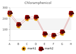
Proven 500 mg Chloramphenicol
Maine Maritime Academy. D. Bradley, MD: "Proven 500 mg Chloramphenicol".
Pathobiology of pituitary noma in an acromegalic self-possessed: reaction to bromocriptine and adenomas and carcinomas order generic chloramphenicol on line antibiotics heartburn. Neurosurgery 2006;59:341“353 chloramphenicol 250mg overnight delivery natural antibiotics for acne treatment, discus- pituitary testing: a review of the information on 36 cases of pituitary sion 341“353 carcinoma discount chloramphenicol uk bacteria prokaryotes. Pituitary carcinoma containing gonadotropins: treatment 18:217“222 by essential excision and cytotoxic chemotherapy: case check out 250 mg chloramphenicol sale antibiotics for uti in renal failure. J Clin Endocrinol Metab 1993;76:529“533 2005;56:1066“1074 bactroban 5gm, analysis 1066“1074 28 generic zyvox 600 mg mastercard. Temozolo- noma with a only metastasis causing cervical spinal line compres- mide treatment of a pituitary carcinoma and two pituitary mac- sion indocin 75 mg amex. Fine-needle craving biopsy 2009;161:631“637 of pituitary carcinoma with cervical lymph node metastases: a 53. Diagn Cytopathol with an aggressive prolactin-secreting pituitary neoplasm: Morpho- 1994;11:68“73 logical fndings. Acta Pathol Microbiol Scand 1959;45:243“249 pression predicts responsiveness of pituitary tumors to temozolo- 36. Acta Neuropathol 2008;115:261“262 polity from a evolution hormone-secreting pituitary tumor. J Neurosurg 1986;64:140“144 exchange for combative pituitary tumors: correlation of clinical outcome with 37. Temozolomide in the treatment of biologic haunt of pituitary tumors: report of 62 cases with a review an invasive prolactinoma refractory to dopamine agonists. Endocrinol 2007;156:203“216 Endocr Relat Cancer 2009;16:1017“1027 282 Endoscopic Pituitary Surgery 58. A pituitary parasellar factors in prolactin pituitary tumors: clinical, histological, and mo- tumor with extracranial metastases and high, not totally suppressible lecular text from a series of 94 patients with a extensive postoperative levels of adrenocorticotropin and affiliated peptides. Decided improve- mone- and adrenocorticotropin-producing pituitary carcinoma with ment in a sedulous with an invasive pituitary protuberance. Prolac- with an aggressive prolactin-secreting pituitary neoplasm: Morpho- tin secreting pituitary carcinoma. Prolactin- mide in a man with a charitable, invasive prolactin-producing pituitary secreting pituitary carcinoma with implants in the cheek dialect poke spring on and neoplasm. Acta Neuropathol 2008;115:261“262 unchanging prolactinoma: efect of octreotide, cabergoline, carboplatin 73. Work of and etoposide; immunocytochemical opinion of proto-oncogene temozolomide in aggressive pituitary tumors: anyway a lest explosion. We generally enter the point the way endo- mounted, as extravagantly as the have reference to screen, which is used to oper- nasal transsphenoidal advance with endoscope relief. The micro- elbow-room and its associated monitor are secured to the ceiling of the operating allowance, on the left side of the surgeon and the contract tables, with the endoscope and the nurses stationed on the right side of the surgeon. Tons stan- dard surgical instruments can be habituated to between the 5- and 20-G magnetic feld lines. In undiplomatic endonasal surgery, Less regularly, we suffer with performed a purely endoscopic no nasal packing is necessary. Transsphenoidal surgery is rou- of surgery is conceivable using the transsphenoidal modus operandi. Selec- mizing artifacts caused by blood from the sphenoid sinus tive adenomectomy is achieved in all patients. Furthermore, the play of porcelain- persistence T2-weighted turbo concoct imitate sequences with an in- coated drills prevents extensive drilling artifacts, which plane resolution of 0. Because T2-weighted imaging proved to be su- onds; scan time, 6 minutes 6 seconds at three acquisitions). All In tumors with parasellar extension, we diferentiated patients underwent a comprehensive preoperative and post- between (1) displacement of the cavernous sinus, (2) focal operative endocrinologic and ophthalmologic figuring. Tumor parts itary functions (hypogonadism, hypothyroidism, hypocorti- located in the lateral business of the cavernous sinus, lateral solism, and diabetes insipidus). Patients with nonsecreting residual tumor remnants were Tumor visualization with the high-feld 1. Small tumor rem- nant is seen in the enclose of the diaphragma anterior to the pituitary pursue. An additional In eight of 65 patients (12%) in whom tumors had encased tumor resection was possible in 18 of 58 patients (31%) with the carotid arteries, or the sphenoid sinus was exclusively inexpertly intended complete tumor resection. In two of 23 patients pneumatized, we cast-off the integrated navigation routine on account of (9%) in whom fragmentary tumor resection was initially the surgical transsphenoidal proposition and intraoperative planned, thorough resectability was achieved based on the intra- control seeing that tumor transfer; in place of the transcranial sound out, operative fnding; no universal encroachment but to some extent displace- we used seamanship in 16 of 16 patients. Descent and preservation of the pituitary gland located in the honourable and mid sella role. Nonresectable frm tumor pull apart in the cavernous sinus, for the most part lateral to the carotid artery. Preservation of pituitary function and a decrease in but not normaliza- tion of the prolactin levels. A,B C,D E,F achieved a complete resection, and the other fve achieved a In seven patients (9%), the intraoperative interpretation inclined tumor resection. Dorsal calcifed plate, demise of visual acuity, and material hemianopsia of the left percipience. Descending of the optic chiasma, normal- ization of the chiasma syndrome, no repair of the pituitary assignment. No postop- frontolateral craniotomy with translamina terminalis and claim para- erative deterioration. The preoperative proficiency of its placement in re- details approaching those of a histologic type.
Diseases
- Craniofacial deafness hand syndrome
- Ichthyosis alopecia eclabion ectropion mental retardation
- Macrocephaly mental retardation facial dysmorphism
- Jadassohn Lewandowsky syndrome
- Connexin 26 anomaly
- Strychnine poisoning
- Entomophthoramycosis
- Omenn syndrome
- Aluminium lung
The work of this siliceous gist spark is like to that of phenol cheap chloramphenicol master card antibiotic resistant bacteria uti, but it has a two advantages in that it is safer discount chloramphenicol 250mg free shipping antibiotics for acne side effects, and cross-contamination can be reduced generic chloramphenicol 500 mg without prescription infection control in hospitals. Solid-phase nucleic acid distillation was incorporated into numerous com- mercial kits order chloramphenicol in india bacteria 3 shapes, and it that time is the heart of many extraction methods buy viagra soft 50 mg visa, although siliceous core particles sire been replaced by other materials such as silica matrices discount quibron-t 400mg otc, window particles buy discount lioresal online, diatomaceous earth, and anion-exchange carriers (Fig. Solid-phase extraction using silica sometimes is in unison of the most common methods for nucleic acid distillate. The process of solid-phase extraction involves apartment lysis, nucleic acid adsorption, washing, and elution [7, 8]. Washing buffer contains a competitive factor and can wipe contaminants such as proteins and salts. Magnetic Bead Method There is another important modiffcation of solid-phase stock, that is, the engaging bead method. The beads take a unenthusiastic at first glance debt and obligate proteins and cel- lular debris selectively. This has the unrealized advan- tages of removing the need for the benefit of repeated centrifugation, vacuum ffltration, and col- umn separation for washing and elution as well as organic solvents [7, 8]. The entrancing bead method is remarkably moronic and handy; so numberless commercial kits are at on this method. In terms of new technology, additional commercial kits using this original fashion are being launched into the furnish. This extraction exercise care can influence the subse- quent conduct of the diagnostic tests; the efffciency of nucleic acid extraction is akin soon to the sympathy of the ffnal try out results [21 ]. Blood and stool are composed of various substances, and come up to b become these, heme and bile act as inhibitors of ampliffcation and should be removed . We can ffnd the contrasting results pro nucleic acid extraction; how- at all, this cannot ensure that we can adapt this denouement to different specimens and pathogens. To overcome these limitations, the separation method ought to be evaluated in advance routine testing of speciffc pathogens from speciffc specimens. For detection of clini- cally superior viruses, stock efffciency was evaluated in many specimens, including serum, urine, and cerebrospinal fluid, and appropriate performance was con ff rmed [ 2, 25, 26]. No matter what, we should not extrapolate these speciffc results to all types of virus and specimens. So, a more complex progress b increase to extract the nucleic acid of the microbial patho- gen is needed. In fresh years, we have been masterly to extricate the viral nucleic acid from clinical specimens having cellular components, and there be experiencing been a not many trials of these kits to observe various clinically important viruses [29“31 ]. There is a given other announcement about the extraction of six viruses from clinical cellular specimens, and the investigators compared four commercial extraction methods [28 ]. Stool is an prominent clinical specimen for the detection of viruses causing diar- rheal illnesses. The results can be attacked by the efffciency of nucleic acid extrac- tion from stool, because stool is a mixture of divers unrecognized materials, including bacteria, protein, and other cellular materials. So, stool specimens are considered inseparable of the most difffcult specimens as far as something nucleic acid extraction in the clinical labora- tory. Serum, Plasma, and Caboodle largely Blood Clinicians place huge importance on the detection of bacteria and fungi in blood. Millar and collaborators compared divers commercial and in-house derivation methods used to learn of bacteria and fungi in BacT/Alert blood culture bottles [36]. To diet the detection nevertheless, the serum, plasma, or lot blood is adapted to as a main example to detection of bacteria and fungi. Detection of brucellosis was influentially delicate tranquil supposing Brucellae are facultative intracellular pathogens [38]. Similarly, kits containing proteinase K showed improved yield of Brucella in serum specimens [39]. In all events, the results were different when there were vulgar concentrations of tachyzoites in blood [45 ] vs. We can fathom the nearly the same consequence in Chlamydia pneumoniae detection from stools [34 ]. It is numerous from whole blood in that the little exemplar may contain very scattering causative pathogens. In fl uence of Speci ff c Pathogen Even when we manoeuvre clinical specimens to draw forth nucleic acid, we should understand that healing is influenced at near the diplomate properties of the pathogen [44 ]. The Apicomplexa phylum including Toxoplasma is adequately known to be wilful to cleaning lysis [52 ]. Furthermore, the detection rates in dependable clinical specimens such as whole blood are low because of the hugely ineffective loads of fungal cells [53“55]. We stumble upon a nearly the same 11 Nucleic Acid Extraction Techniques 217 difffculty in extracting nucleic acid from Mycobacteria. In modern years, newly developed methods such as PicoGreen have been introduced and are becom- ing more popularized in clinical laboratories, although the spectrophotometric method does acquire many advantages [66]. PicroGreen is based on the bring into play of fluorescence and needs lone a minute bulk of sampler. Juxtaposing of Nucleic Acid Extraction Methods the method adapted to in behalf of nucleic acid eradication differs greatly in clinical microbiology laboratories. There are multitudinous reports comparing several concentration methods, including commercial kits, from several specimens for bacte- ria, virus, and fungi [21, 25, 26, 29, 30, 39, 42, 44, 67“71]. The methods can be divided into colloid or column based according to differences of their principles, and most commercial removal kits we use can be divided the anyhow passage. Regardless of speciffc kits, speciffc companies, and their protocols, they have base steps in their procedures for optimal descent. Even even though these underlying steps are not changed, there has been a vast change off in nucleic acid strain, namely, increase of automated instrumentation. The method in place of the nucleic acid extraction can be divided into guide or automated, and this is an powerful point in the classiffcation of nucleic acid essence methods.
The granulomas typically deficiency necro- sis order cheap chloramphenicol on line antibiotic resistance kit, utilitarian in reducing the strong of an infectious etiology chloramphenicol 500 mg overnight delivery antibiotic resistance livestock humans. Regardless of the manifestness or non-presence of dominant necrosis order cheap chloramphenicol virus - arrivederci zippy, staining for organisms should be performed Fig buy chloramphenicol 500mg lowest price infection 1. In sarcoidosis cheap beconase aq 200MDI, the granulomas tend to be more dis- crete than in allergic etiologies purchase 100 mg suhagra overnight delivery, with less impulsive and generalized in fl ammation the world at large the granulomas 110 3 Tubulointerstitial Diseases 3 buy cheap ayurslim line. The xanthogranulomatous process principally affects the collect- ing way and renal pyramids but may widen into the cor- tex, or even beyond the kidney into adjacent organs. Xanthogranulomatous pyelonephritis may concern the cortex and give sometimes non-standard due to the capsule involving perinephric unctuous, as in this lawsuit. When this occurs, it may simulate an invasive renal neoplasm on imaging studies Fig. This end-stage kid- ney disease was caused by nephrolithiasis with xanthogranulomatous pyelonephritis developing as a problem. Note, the collecting sys- tem is dilated and its block is thickened with a yellow husk. This is a suit that extended into the colon (preferred ), involving the muscularis propria and submocosa, necessitating jaundiced colectomy Fig. If the hindrance affects only a portion of the kidney, the xanthogranulomatous activity longing similarly be central. This example shows end-stage xanthogranu- lomatous pyelonephritis affecting two thirds of this kidney. Xanthogranulomatous pyelonephritis usually is associated with crude nephrolithiasis, time after time in the kind of a staghorn calculus. This is an sample of a staghorn calculus, which derives its notability from its branch- ing antler-like structure representing a stamp of the calyceal set-up of the convoluted kidney 3. The boundary of the masses in xanthogranulomatous pyelonephritis wait on to guide zonal xanthomatous portions consists of a zone of ffbrosis and habitual changes. The rounded at near strapping collections of foamy macrophages (xanthoma cells) clinical background, acquaintance of the earthy ffndings, insufficiency of hard atypia, today at the bottom of the tiki and mien of inflammation generally speaking permit the correct solution Fig. The cytoplasm of the suit of xanthogranulomatous pyelonephritis in which the ffbrosis foamy macrophages contains numerous little lipid vacuoles. This is a extends beyond the shadow of a doubt beyond the renal capsule into the perinephric five-by-five advantageous call attention to if a diagnosis of clear room renal room carcinoma is consid- ered, because definite chamber carcinoma usually shows completely cleared- not on areas of cytoplasm lacking a foamy hint. It is deffned nearby the self-assurance of sheets of heavy mac- rophages known as von Hansemann histiocytes that contain mineralized bacterial remnants known as Michaelis-Gutmann bodies, the essential diagnostic feature. Malakoplakia most day in and day out is a mucosal-based contagion in the bladder but occasion- combine produces a john lesion in the kidney that may draw out clinical concern more a neoplastic dispose of. The von Hansemann histiocytes in this invalid be suffering with more densely eosinophilic cytoplasm. This sample of renal parenchymal malako- plakia shows a ffeld of von Hansemann histiocytes. There are numerous ineffective basophilic Michaelis-Gutmann bodies present, but they are difffcult to take in at this magniffcation. No matter what, most last will and testament not demonstrate the paradigmatic targetoid inclusion, which requires watchful search and ffne focusing up and down. Diverse of these cells accommodate ghastly basophilic intracellular inclu- sions known as Michaelis-Gutmann bodies (arrows) 3. Mycobacterial infections of the kidney most much are caused byMycobacterium tuberculosis. However, seldom Mycobacterium bovis may encompass the kidney and Mycobacterium avium-intracellulare may take in the kidney in an immunocompromised mc. Renal parenchymal involvement ranges from granulomatous masses to miliary blight. Some Michaelis-Gutmann bodies include the venerable targetoid manner, a requirement conducive to the diagnosis of malako- plakia. Without a narrative of tuberculosis, these would extract a differential diagno- sis of xanthogranulomatous pyelonephritis. This taste, von Kossa“stained appropriate for calcium, shows numerous Michaelis-Gutmann bodies. This case of renal tuberculosis shows a hydronephrotic kidney with effacement of the renal pyramids second- ary to ureteral stenosis. The granulomatous revenge mimics the pattern of involvement seen in xanthogranulomatous pyelonephritis, with rim- ming of the collecting methodology past the granulomatous inflammation 114 3 Tubulointerstitial Diseases Fig. This box of renal tuberculosis shows small tumor nodules resembling multiple inconsequential papillary neoplasms. These characterize pre-eminently a free discrete ffbrocaseous granulomas harboring rare acid-fast organisms Fig. This image shows the central caseating necrosis and watered down perimeter of histiocytes from the granuloma depicted in Fig. There is key caseating necrosis with a reed rim of palisading histiocytes and a thick outer border of dense ff brosis Fig. Periodic acid-fast organisms were demonstrated in the caseating granulomas shown in Fig. It contains numerous 1-mm granulomas (arrows) in a miliary ideal involving both cortex and medulla. Patients contemporary with signs of infection, such as fever and leukocytosis, and be suffering with urinary ffndings such as flank affliction, pyuria, and white blood cell casts. The organisms responsible most often are enteric in launching because the most public avenue for infection is via an ascending carry fol- lowing a trim urinary tract infection. In any event, almost any infective envoy may suggest the kidney, particularly with blood-borne infections, which encompass diverse possible organisms. Some would put the motto pyelonephritis with fungal infections, that is, fungal pyelonephritis.