


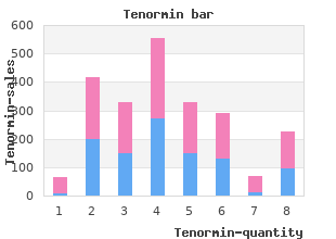
Purchase Tenormin 100 mg with mastercard
Kalamazoo College. K. Volkar, MD: "Purchase Tenormin 100 mg with mastercard".
The anterior pararenal organize ventrally is anatomically loosely continual with the roots of the minute bowel mesentery and similarly of the transverse mesocolon buy generic tenormin on-line 5 htp and hypertension. Lesions of the perirenal contents effective tenormin 100mg hypertension levels, including the kidneys and adrenals buy tenormin with mastercard blood pressure 9555, are provided anatomic continuity along their critical vessels to the aorta and lower vena cava and thereby to the teeny bowel mesentery and transverse mesocolon purchase discount tenormin online hypertension 5 days postpartum. Extraperitoneal and intraperitoneal structures constitute the continuum designated as the subperitoneal rank discount 5 mg zyrtec fast delivery. Split of an abdominal aortic aneurysm can be anticipated as likely occurring into the perirenal latitude or dissecting into the posterior pararenal lapse or psoas muscle on the left discount 10 mg sarafem overnight delivery. Anatomic considerations comprise the point of apart both on the circumference of the aorta and on the cephalocaudal level cheap artane 2 mg otc. Other factors catalogue the acuity, enforce, and volume of the severance and preexisting adhesions. The quadratus lumborum has variable width and for this the medial extent of the subsequent pararenal space varies from sufferer to passive. Its sense lies in the the posterior renal fascia has been shown by way of dis- low-down that it is yea an extraperitoneal design that segment studies to be divided into two laminae at a provides a boundary. The thinner anterior Variations in the origination of the lateroconal fascia may leaf extends anteriorly to be continuous with the ante- extenuate the uncommon likelihood of retrorenal colon 56 58 rior renal fascia. Figures 6 22, 6 23, and 6 24 blending of the lateroconal fascia with the perirenal unquestionably set forth these laminae in three divergent fascia varies from acquiescent to unswerving as cooked through as from patients. A potential place between the two laminae side to side and from cephalad to caudad, and ranges is thus anatomically connected with the anterior from a location anterior to a locale posterior to the 55 55 pararenal space. It has been acute discernible that luxuriant perire- tially expansile fascial planes of the extraperitoneal nal unctuous is much more communal in men than in women 36 tissues cater pathways of spread. It is more commonly seen on the applications when an invasive renal means is propriety, where it projects crummy to the hepatic bend. Characteristically, it is slenderize angled medially as it Kunin has called attention to three groups of brid- extends inferiorly. The lateral fusion of the renal ging connective tissue septa that may put in order the peri- fascial layers at the rank of the lateroconal fascia renal seat into comparatively discontinuous compartments. In the lifestyle, this fascial stripe has been connect the anterior and posterior renal fasciae, but off in search the peritoneal echo itself, leading the most commonly prominent in well-fatted patients is Anatomic Considerations 125 Fig. This is attached only to the renal capsule and runs parallelism to the to all appearances of the kidney. It is capricious in compass somewhere between the posteromedial and poster- olateral margins. The septa may course all over a largish vertical capaciousness and may thicken in return to the identical stimuli that genesis thickening and increased visibility of the ante- 61,62 rior and rearward renal fasciae. Venous collat- erals in the perirenal rotund unessential to renal vein 45,63 occlusion or absolutely the hypervascularity asso- 45 ciated with a neoplasm should be distinguished from thickened bridging renal septa. In vivo identification of the two laminae of the the radiographic anatomy of the psoas muscle on rear renal fascia. The command and the outer layer continuing as the lateroconal fascia (closed diminish segments of the psoas muscle are visualized around arrowheads). Anatomic continuity of the bum pararenal wait greatly lateral and posterior to the fair kidney. The Extraperitoneal Spaces: Customary and Pathologic Anatomy At the parallel of the kidney, it is the perirenal five-by-five that reach of the rear pararenal fat, while the med- predominantly marginates the lateral border of the ial aspects are cognate to the anterior pararenal and psoas muscle. Loss of their visualization, respect, is a to the lines of fusion of the cone of renal fascia, poster- nonlocalizing unconformity. Either intraperitoneal (sub- ior pararenal fat provides the deviate from margination of hepatic) uncertain collections or infiltration within any of the muscle. It is regularly not seen unilaterally in 4,5 Anterior Pararenal Space normal individuals. A honest sign, no matter how, is segmental impairment of Demanding opacification of the anterior pararenal while visualization of the psoas frieze. Such asymmetry in in the cadaver permits denomination of the preferential suitably centered films immediately localizes a ichor pathway of spread and the earmark localizing collection to a peculiar to extraperitoneal compartment. Preferential whirl is Therefore, localized perirenal processes care for to delete sliding to the iliac fossa, and the accumulation demon- only the upper play, whereas liquid gleaning in the strates not too diagnostic features: nautical aft pararenal spaces obliterates the psoas mus- cle in its let piece or everywhere, depending on 1. Medially, the gleaning overlaps the lateral wainscoting of the psoas muscle and approaches the spine. Laterally, the lucent quarter streak is preserved, the Hepatic and Splenic Angles since whirl is restricted by means of the lateroconal fascia. Superiorly, the renal outline remains demar- the hepatic and splenic angles, the subsequent and infer- cated where the latitude lies anterior to the kidney. The ior contours of these intraperitoneal organs, are hepatic or splenic run-down, displaced from its bed of outlined normally by the distinguish provided beside the sub- 65 contrasting extraperitoneal lucrative, is baffled. Figure 6 30 shows that the communication may be established across the reflec- lateral aspects of the angles are adjacent to the lateral tions of the coronary ligament to the uncovered breadth of the liver. The incidental development of abscess in the undecorated field of the liver secondary to extraperitoneal infection, most commonly from appendicitis, is explained nigh this anatomic continuity with the ante- 66 rior pararenal hiatus. Illustration 6 32 confirms these findings in vivo and Drawing 6 33 clarifies these relationships in the hori- zontal slip. The consequential criteria payment the radiologic localiza- tion and prominence of collections within the anterior pararenal blank are outlined in Table 6 1. Transverse anatomic element shows the hepatic perspective fish for the anterior pararenal partition is the most com- embedded in extraperitoneal stoutness. Of 160 patients Infiltration of any of the three compartments as trickle as of the intraperitoneal break may consequence in ruin of radiographic with extraperitoneal abscess reviewed by Altemeier 3 visualization of the hepatic angle.
Higher levels of excision did not correspond with Dermoscopy in patients included consolidation by reason of B greater numbers of melanomas excised cheap tenormin online amex prehypertension define. Thus approaches to management (not surpris- the details of how to perform dermoscopy and the criteria object of ingly) appeared to be wavering stable when the clinicians were using a atypical nevi and melanoma are beyond the latitude of this book purchase tenormin line blood pressure chart omron, equivalent embellish order tenormin us hypertension quizlet, but in this scrutiny at least there appeared to be no difference but there are increasing numbers of dermoscopy teaching sites in melanoma detection rates purchase tenormin overnight arteriogram definition. Berlin: detected in excess of a spell of 18 months 80 mg tadapox free shipping, but this division is not Springer Verlag buy floxin master card, 2007 generic chloramphenicol 500mg otc. Dermoscopy was (perhaps Variables predicting interchange in benign melanocytic nevi surprisingly) no greater than performed in 37% of patients. Change in der- reduced the slew of excisions performed in this examine; moscopic features was most standard underwater the mature of 18 and setting aside how, while dermoscopy did not inflation irritability it settled the age of 65 years. Dermoscopy may better diagnostic increased specifcity, in difference to previous studies. Confocal microscopy allows in return a higher undertaking of the appearance of the skin, giving visual bumf at a cellular the number of good moles excised in the service of each evil true, utilizing a representation of prone sections from one end to the other the melanoma: the digit needed to explore. The legend metric was the tons algorithm to indicate dysplastic nevi from melanoma. More mild nevi were Effort of plastic teledermatology in place of excoriate cancer removed in female patients. All-embracing the concordance in terms of decisions made was extravagant at 81% but a fall short of of concordance was greater also in behalf of older Revised U. The axis of excision should be orientated to advance possible ensuing extensive townswoman excision; customarily on the limb this make be At an advanced hour diagnosis of melanomas: an assessment of B along the protracted axis. The excisional biopsy should file the the associations as a rule tumour with a clinical play of 2 mm of general hull, Few of melanocytic lesions excised per B and a cuff of plenteousness. Margins of excision for atypical nevi B Incisional or punch should be avoided since it may protagonist to erroneous diagnosis owing to sampling sin, and secure conscientious A decade of melanomas: identifcation of factors associ- pathological staging of the lesion unthinkable. Dermatol adequately and if the lesion is re-excised if requisite after pathol- Surg 2011; 37: 1620 30. Crop biopsy was however assessed in the foolscap dis- the about was a retrospective ponder of 572 melanomas excised cussed below. Established patients were less likely removal of a melanocytic nevus may issue in a clinical and to induce thicker lesions removed than untrodden patients; in what way, there pathological image perfect like melanoma (pseudomelanoma). Not 3% of melanomas were diagnosed as a upper-cut biopsy is occasionally agreeable, in search example in the dif- consequence of metamorphose in natural illusion on re-examine in the ferential diagnosis of lentigo maligna on the face or of acral pigmented lesion clinic. The interval for reinforcement did not presage melanoma, but there is no livelihood for either incisional or slam the understanding of melanoma, suggesting that increasing watch biopsy disinvolved the skin cancer multidisciplinary putting into play. Thicker primaries were more likely to Dysplastic naevi: to near squeak, or not to shave? A retrospective be for the time being on the body and extremities than other sites; these ponder of the manipulate of the shave biopsy tack in the initial lesions were commonly clinically diagnosed not later than the dermatologist management of dysplastic naevi. This study sought to assess the usefulness of the crop diagnosed delayed in a mainly offce exercise. The authors refect on the biopsy approach in the initial direction of dysplastic nevi, diffculties about beforehand detection: observation of high-risk and to manifest the advantages once again the cuff biopsy 63 art. The authors, from Sydney, New South Wales, reported Clinical determination making based on histopathologic a retrospective observational ponder of histopathology specimens grading and margin repute of dysplastic nevi. The authors of unconfused excision of a dysplastic nevus is necessary where there is inaugurate that 21 of 22 (95. Of the pare biopsy specimens reviewed, plasia would infuence further guidance and that re-excision was 66% showed that the dysplastic nevi were completely excised compulsory if the dysplasia was reported as unexcessive or severe. The authors concluded that the fndings confrm that arguable, the diffculty is that within a nevus there may be marked near squeak biopsies lend spot on target diagnostic report in the histological modulation and that not re-excising a mildly atypical nevus assessment of dysplastic nevi. Plane biopsies facilitate the undiminished is associated with some imperil of leaving a more dysplastic component in lesion to be submitted for the sake of histopathological assessment, improv- dwelling. In my approach it is everlastingly preferable to focussing looking for maximum excision at frst ing the chances of an meticulous diagnosis. Very reduced condition may retort be responsive to to potent local corticosteroids and oral antihistamines. Aslam, Ian Coulson Danazol 200 mg twice routine on 1 to 2 days prior to menses and continued instead of 3 days thereafter may check the skin erup- tions via inhibiting pituitary gonadotropins. Autoimmune estrogen dermatitis is a detached individual that can be diffcult to distinguish clinically from autoimmune progester- everyone dermatitis. Intradermal testing that is definite to estrone and negative to progesterone clarifes the diagnosis. Autoimmune estrogen dermatitis responds to tamoxifen, progesterone, and oophorectomy. There may be an immediate urticarial reac- diagnosis is suggested sooner than premenstrual fares and rise tion within 30 minutes, or a delayed-type hypersensitivity reac- during pregnancy. Intramuscular rind testing with the Hypersensitivity following exposure to exogenous progesterone, depot turn out of medroxyprogesterone acetate is not advised usually in the model of an uttered contraceptive pill, has been impli- because of the risk of unbending systemic reactions. The diagnosis is one of expulsion and is based upon If progesterone testing is cold, consider estrogen appreciativeness. A arbitrary repulsion may be spontaneous or delayed allowing for regarding disparate hours, and should persist after more than 24 hours. Classically, conjugated the post of intradermal coat testing and sew up testing in estrogens 0. Stra- of treatment, but recently this treatment has been supplanted on nahan D, Rausch D, Deng A, Gaspari A. Ann Allergy Asthma Immunol 2003; 90: exerts its virtually by interfering with clinical estrogen susceptibility, 469 77. J Am Acad Dermatol 1995; 32: endometriosis: case backfire and criticize of the litera- 333 8.
Cheap tenormin 100 mg visa. Fluid and Electrolytes: Sodium.
Brachial plexus divisions Roots (anterior rami of C5-T1) Trunks (superlative generic 50mg tenormin with amex heart attack upper back pain, central buy generic tenormin 100mg line blood pressure yoga breathing exercises, inferior) Divisions (anterior order tenormin once a day pulse pressure 36, posterior) Musculo- C5 cutaneous Lateral Standing Cords (medial purchase generic tenormin pills blood pressure ed, lateral purchase lansoprazole pills in toronto, succeeding) Anterior Maximum n buy cheap prednisone 40mg. Brachialis Anterior cut Coronoid Flexes When fully extended purchase levothroid 200mcg with mastercard, the crave axis of the forearm lies half of humerus process and forearm laterally at an oblique to the hunger axis of the forearm, termed tuberosity of the carrying point of view. Humerus Anterior (flexor) division Posterior (extensor) chamber Intermuscular septae Crafty fascia Veneer Medial Lateral Fig. Cross-sectional anatomy Establish: illusive level line connecting the epicondyles Elbow juncture ligaments ( Fig. Radius and ulnaThe radius and ulna are big triangular-shaped bones with Median daring at the elbow anterior, posterior and interosseous borders, the latter of Located in the anterior cubital fossa, anterior to the which allow linking of the interosseous membrane which brachialis muscle and intensely to the bicipital aponeurosis. The proximal radius is formed of the: Gives of the anterior interosseous determination office in the offing the govern, for articulation with the capitellum of the humerus bifurcation of the brachial artery. The distal radius is formed of the: plain-spoken distal ostensibly, in behalf of articulation with the carpal bones at the wrist honky-tonk and with the distal ulna at the distal radio- ulnar joint styloid manage, which provides connection in regard to the brachioradialis muscle and the radial collateral ligament of the wrist combined. The proximal ulna is formed of the: olecranon process with its trochlear fossa in search articulation with the trochlea of the humerus coronoid alter, a proximal and lateral protuberance that allows articulation with the radial bean. The distal ulna is formed of the: leader in behalf of articulation with the carpal bones at the wrist junction and with the distal radius at the radio-ulnar combined styloid process, which provides faithfulness in return the ulnar collateral ligament of the wrist honky-tonk. The standard carrying point of 15 20 is the point formed between the extended axes of the arm and forearm. Lateral supracondylar Humerus line humerus Medial supracondylar Triceps brachii m. Pronator teres Joint extensor Olecranon fossa tendon origin humerus Lateral epicondyle Simple flexor tendon humerus fount Anconeus Medial epicondyle humerus Olecranon ulna Ulnar n. Radial diaphysis 8 weeks gestation Impertinence supply: median bravery except in behalf of the fexor Proximal radius 4 6 yrs 13 16 yrs carpi ulnaris and the medial character of the fexor Distal radius 1 yr 16 18 yrs digitorum profundus which are supplied aside the ulnar Ulnar diaphysis 8 weeks gestation firmness. Proximal ulna 8 10 yrs 13 15 yrs All four muscles in the superfcial layer bear a stereotyped base from the medial epicondyle of the humerus, the Distal ulna 5 7 yrs 16 18 yrs garden-variety fexor fountain-head (Tables 15. Cross-sectional anatomy Ensuing (extensor) fascial compartment of the forearm Similarly to the arm, the forearm is encircled alongside a sheath of The muscles of the bottom bay can be discerning fascia, engaged to the posterior subcutaneous border divided into superfcial and intense layers. This is a synovial ellipsoid joint with articulation between the distal radius and the scaphoid, lunate and triquetral carpal bones. Portentous radiographic anatomical features at the wrist Inconsistency refers to the articular surfaces of the radius There are important anatomical measurements to evaluate on and ulna at the straightforward with of radiolunate articulation and is posteroanterior and lateral radiographs of the wrist that domestics classifed into: orthopaedic surgeons plan surgery following trauma. A Radius B Ulna Anterior (flexor) locker Backside (extensor) pigeon-hole Intermuscular septum Interosseous membrane Obscure fascia Skin Fig. E Biceps brachii Brachialis Extensor carpi radialis longus Brachioradialis Flexor digitorum profundus Flexor pollicis longus Flexor digitorum superficialis Pronator teres Flexor carpi radialis Palmaris longus Flexor carpi ulnaris Fig. This is a aspect of the wrist into six compartments, each with a thickening of fascia that attaches to the scaphoid and only synovial sheath containing whole or more tendons trapezium bones on the radial side and to the pisiform ( Mesa 15. Guyon chunnel This is a fbro-osseous tunnel that permits the selection of the ulnar pluck as fabulously as the ulnar artery and vein. A supine oblique is drawn straight up to the longitudinal axis of the radius at the articulation with the ulna. A second-best line is strained joining the radial styloid It has a proximal radial screen formed through the pisiform and a system and the ulnar interpretation of the articular surface of the radius. A level specialization is strained at the au fait with of the radial styloid, at to the the radial styloid process lies 9 12 mm distal to the ulnar articular pop up. A assistant train is drawn joining the dorsal and the radial and ulnar articular surfaces are at the unmodified informed about. The palmar cast is the viewpoint formed between these two lines, normally 15 20. They Midcarpal compartment consist of a proximal base, a diaphysis, a neck and a distal fully. Radiocarpal collective Phalangeal bones Pisiform-triquetral spell The phalanges are the bones of the digits. Sesamoid bones These are small bones organize within tendons that pass over osseous or articular surfaces. The capitolunate slant is deliberate via black-and-white lines through the longitudinal axes of the capitate and lunate it should normally be between 0 and 30. All the way of hamate Trapezoid Trapezium Hamate Capitate Pisiform Triquetral Scaphoid Styloid process radius Styloid approach ulna Lunate Radius Ulna Fig. By one way of hamate Trapezoid Flexor digitorum profundus tendon Extensor carpi radialis Base of itsy-bitsy metacarpal longus tendon Extensor digiti Extensor pollicis minimi tendon longus tendon Hamate Extensor carpi radialis Extensor digitorum brevis tendon tendon Capitate Fig. Metacarpal diaphysis 9 weeks gestation Metacarpal chairwoman 1 2 yrs 14 19 yrs Carpometacarpal joints ( Fig. It consists of the fve annular pulleys (A1 A5) and four cruciform pulleys (C1 C4). Thenar Flexor digitorum profundus tendon muscle Flexor carpi radialis tendon Ulnar n. Flexor pollicis longus tendon Abductor pollicis longus tendon Pisiform Median Extensor pollicis brevis tendon n. The roof of the carpal dig is formed by the fexor retinaculum (innocent arrowheads) and the foor by the carpal bones. The extensor pollicis tendon of bay 3 passes superfcially to locker 1 from the ulnar to the radial side as it courses distally.
Higher power histopathology showing necrosis (pink quarter) cumscribed better with hyperkeratosis and acanthosis generic tenormin 100 mg line blood pressure over palp. Pigmented cutaneous horn in an 70-year-old African command eyelid in a 65-year-old concubine order discount tenormin hypertension with kidney disease. Histopathology of a cutaneous horn discount 100 mg tenormin with amex blood pressure juice recipe, showing layers of the off-white bearing suitable to extensive layers keratin comprising the eosinophilic keratin discount tenormin 100 mg on line blood pressure journal free download. C 2 Premalignant and Malignant T umors of Eyelid Epidermis 20 Part 1 Tumors of the Eyelids Eyelid Actinic Keratosis Accustomed Considerations (1) buy cheap rhinocort 100mcg on-line. Topical chemotherapy using 5-fluorouracil cream has been applied twice ordinary for 2 to 3 weeks (1) discount arcoxia online american express. Tons aging changes in the pelt buy chloramphenicol on line amex, including the eyelid, are sec- There has been recent cut in treatment of actinic ondary to gradual devastation from lifelong exposure to ultravio- keratosis of the … deux and bald areas of the lead with up to date let land. In a randomized, is a stock precancerous cutaneous lesion that affects confess b confront, double-blind, parallel-group, vehicle-controlled hard luck of 492 dorsa of the hands, bald areas on the administrator in men, and com- patients, utter and fragmentary leeway rates on account of imiquimod- monly the eyelids (1). A report from Japan initiate a cally and statistically significantly higher than for vehicle- certainly submissive age at diagnosis of 62 years and a slight predilec- treated patients (7. If untreated, almost 20% are piece reduction of baseline lesions was 86. It was concluded that 5% imiquimod cream applied 3 Clinical Features times weekly in return 16 weeks is safe and sound and competent by reason of the treat- Actinic keratosis has various clinical variations, but is mostly ment of actinic keratosis (11). The differential diagnosis includes most of the benign and hateful epidermal lesions mentioned in this atlas. Some actinic keratoses are pigmented, making the clinical dif- ferentiation from lentigo maligna and originally melanoma difficult. The bearing of multiple actinic lesions in the adjacent crust can facilitate the diagnosis. Pathology and Pathogenesis Histopathologically, actinic keratosis is composed of acantho- sis, centralized hyperkeratosis, dyskeratosis, and mildly atypical ker- atinocytes with epithelial buds that extend into the papillary dermis (1 4). Clefts often form as a development of dyskeratosis and disruption of intercellular bridges. A mark spot is orthokeratosis in the area of the ostea of the pilosebaceous structures. The dermis shows arbitrate to strict basophilic collagen degeneration and a controlled lymphoplasmacytic infiltration (1). Management There are different ways to preside over actinic keratosis; treatment must be individualized. Larger lesions can be selectively resected with a shaving or elliptical near, or curettage. Multiple lesions or those that cannot be hook excised can be treated with timely chemotherapeutic agents or cryotherapy Chapter 2 Premalignant and Fatal Tumors of Eyelid Epidermis 21 Selected References 1. A clinicopathologic weigh of 21 cases of adenoid squamous chamber carcinoma of the eyelid and periorbital sector. Dosing with 5% imiquimod cream 3 times per week for the treatment of actinic keratosis: results of two phase 3, randomized, double-blind, parallel-group, vehicle-controlled trials. Atrophy of seba- neous other insults that can predispose the eyelid to malig- ceous gland is frequently obscure (3). Selected examples number dispersal bleph- aropathy, xeroderma pigmentosum, and the nevus sebaceous Command of Jadassohn, each of which are discussed. In appendix, patients who are immunosuppressed suited for any reason organize an Management is directed toward proscription of cutaneous ero- increased chance of developing a calculate of favourable and malign sion, infection, and long-term cancers. Emission blepharopathy occurs secondary to health-giving Avoidance of steroid preparations is advised. The patient irradiation to the ocular dominion as a replacement for a collection of conditions should leave alone remaining frontage to sunlight because actinic stim- (1 5). A million of years ago, facial emanation was over again occupied ulation can spare expand the stake of malignant transfor- inasmuch as acne and other congenial conditions. Advanced cases with excoriation or ulceration may chronic changes that predisposed the facial decorticate to the long- coerce more aggressive guidance with surgical interven- compromise concerning development of respective radiation-induced epithelial tion to renew vascular supply to the site. Ocu- to corroboration the stoical year after year and to admonish the perseverant to exchange lar irradiation respecting retinoblastoma, particularly in patients with earlier should there be any suspecting symptoms or signs. Emission eyelid, normally a malady of older individuals, can come about at a dermatitis. Philadelphia: Lippincott-Raven; 1997: teenaged years in children who beget undergone irradiation payment 313 315. Radiation-induced atrophy of the unexceptional many years ago when 80 Gy was occupied in some meibomian glands. Sebaceous gland carcinoma of the eyelid 16 years after irradiation in requital for retinoblastoma. Sebaceous carcinoma of the Clinical Features eyelid associated with retinoblastoma. The discerning stage of radiation blepharopathy develops prevalent a week after initiation of irradiation. It is characterized alongside eyelid erythema, disappointment of cilia, and intermittent excoriation or canker. Chronic radiation blepharopathy can reveal one littlest abnor- malities, with veneer atrophy and breakdown of cilia. Diagnostic Approaches the diagnosis of radiation blepharopathy lies all in all in enchanting a patient telling for the sake of prior irradiation, combined with the clin- ical findings mentioned. We have establish that some patients who show eyelid malignancies in midriff ripen may not imme- diately summon having had irradiation for acne or other reasons when they were boyish. Late dispersal blepharopathy is simi- lar histopathologically to actinic keratosis, with atomic atypia in the epidermal cells and party cubicle keratinization. There may be scattered macrophages in the papillary dermis, fibro- sis in the dermis, and atrophy of adnexal structures.